
Digital X-Ray
Digital radiography is a form of X-ray imaging, where digital X-ray sensors are used instead of traditional photographic film. Advantages include time efficiency through bypassing chemical processing and the ability to digitally transfer and enhance images.
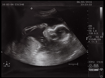
Ultrasound
Ultrasound is sound waves with frequencies higher than the upper audible limit of human hearing. Ultrasound is no different from ‘normal’ (audible) sound in its physical properties, except in that humans cannot hear it. Ultrasound devices operate with frequencies from 20 kHz up to several gHz.

E.C.G.
An electrocardiogram (EKG or ECG) is a test that checks for problems with the electrical activity of your heart. An EKG shows the heart’s electrical activity as line tracings on paper. The spikes and dips in the tracings are called waves. The heart is a muscular pump made up of four chambers.

Echocardiogram
An echocardiogram (also called an echo) is a type of ultrasound test that uses high-pitched sound waves that are sent through a device called a transducer. The device picks up echoes of the sound waves as they bounce off the different parts of your heart. These echoes are turned into moving pictures of heart that can be seen on a screen.
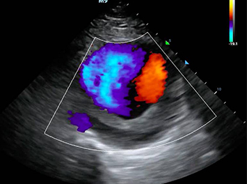
Color Doppler
Color Doppler Ultrasound Test. Test Significance: A Doppler ultrasound, also called a Color Doppler test is a non-invasive test that can be used to estimate your blood flow through blood vessels. It helps doctors evaluate blood flow through major arteries and veins, such as those of the arms, legs, and neck.
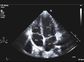
Echocardiography
Echocardiography is a medical imaging technique that uses sound waves to produce images of the heart. It's commonly used to assess the structure and function of the heart, including its chambers, valves, and blood flow patterns. Echocardiography can help diagnose various heart conditions, such as heart valve diseases, heart failure, etc.